.jpg "Elms_hub.jpg")
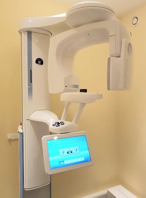 We are delighted with our new Cone Beam Computed Tomography (CBCT) scanner, state-of-the-art technology producing three-dimensional images of your teeth, soft tissues, nerve pathways and bone in a single scan.
With CBCT, an x-ray beam in the shape of a cone is moved around the patient to produce a large number of high-quality images. In a single rotation, the detector can generate anywhere between 150 to 200 high-resolution two-dimensional images, which are then digitally combined to form a three-dimensional image that can provide your dentist or oral surgeon with valuable information about your oral and craniofacial health. This new technology uses ultra-low-dose radiation to obtain exceptional images.
Three-dimensional images obtained with CBCT allow for more precise treatment planning, such as:
A CBCT examination requires no special preparation. Prior to the examination, you may be asked to remove anything that may interfere with the imaging, including metal objects, such as jewellery, glasses, hairpins and hearing aids. Although removable dental work may need to be removed, it is advisable to bring these to your examination, as your dentist or oral surgeon may need to examine these as well. Women should always inform their dentist or oral surgeon if they are pregnant.
Your dentist will position you so that the area of interest is centred in the beam. You will be asked to remain very still while the x-ray source and detector revolve around you for a 360-degree rotation or less. This typically can take between 20 to 40 seconds for a complete volume, also called a full mouth x-ray, in which the entire mouth and dental structures are imaged, and less than 10 seconds for a regional scan that focuses on a specific area of your jaw.
Your images will be combined to form a three-dimensional image, which can be shown directly to you back in the comfort of your dentist's surgery. Your dentist will discuss the findings and make any recommendations on treatment based on these results.

With fast scanning times, low radiation doses and a range of fields of view and resolutions, CBCT offers exceptional insight for treatments such as dental implants, endodontics (root canal treatment), orthodontics (teeth straightening) and oral surgery.
Lead nurse Kriszti and dentists Kit Spears and Hugh Cowley receiving certification for our #Planmeca ProMax Classic 3D CBCT scanner from Project Manager & BDM Chaz Sidhu, installed by #Blueprintdental with demo support and Applications Training delivered by Jonathan Hampton #Planmeca.
